Introduction
Solid Pseudopapillary Neoplasm (SPN), also known as Frantz tumor, is a rare pancreatic tumor first described by Frantz in 1959 [1]. It was formally recognized as a distinct clinicopathologic entity by the World Health Organization (WHO) in 1996 [2].
SPNs represent approximately 1–2% of all pancreatic tumors [3-5]. They predominantly affect young females in their second or third decade of life, with a female-to-male ratio of approximately 10–11:1 [3,4].
Although SPNs are classified as low-grade malignant neoplasms of the exocrine pancreas, they exhibit indolent behavior and favorable prognosis. Since 2000, the reported incidence has increased nearly sevenfold, likely due to improvements in imaging modalities and heightened clinical awareness [6].
Approximately 60% of SPNs arise in the pancreatic body or tail, while the remainder occur in the head [3,7]. Up to 70% of cases are asymptomatic and discovered incidentally [7]. When symptomatic, patients typically present with nonspecific abdominal pain.
Serum tumor markers (AFP, CEA, CA19-9, CA125) are generally within normal ranges and lack diagnostic specificity [8].
Surgical resection with negative margins remains the cornerstone of treatment and offers cure rates exceeding 95% in localized disease [3,4].
This report describes a rare case of SPN in a 14-year-old girl and reviews the relevant literature.
Case presentation
A 14-year-old previously healthy girl presented with intermittent abdominal pain of several months’ duration. There was no history of weight loss, jaundice, fever, or prior abdominal surgery.
On examination, she was stable with no palpable abdominal mass. Laboratory investigations, including liver function tests and tumor markers (AFP, CEA, CA19-9), were within normal limits.
Investigations
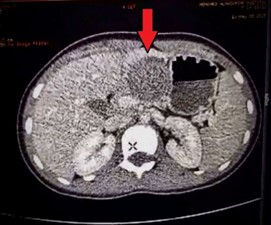
Contrast-enhanced Computed Tomography (CT) demonstrated a 2.5 cm, well-defined mass located in the pancreatic body and tail. The lesion exhibited mixed solid and cystic components with peripheral calcifications. No vascular invasion, lymphadenopathy, or distant metastases were observed (Figure 1).
Magnetic Resonance Imaging (MRI) showed:
• Hypointense signal on T1-weighted images
• Hyperintense signal on T2-weighted images
• Well-encapsulated margins without surrounding infiltration Based on radiological characteristics, a presumptive diagnosis of SPN was made. Preoperative biopsy was not performed due to characteristic imaging features and resectability.
Surgical management
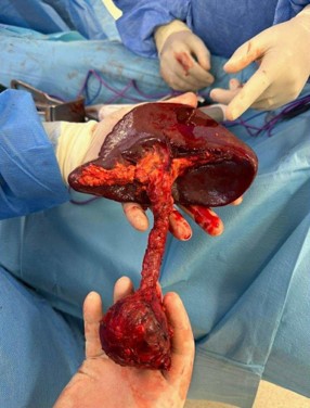
The patient underwent distal pancreatectomy with splenectomy. Intraoperatively, the tumor was encapsulated and confined to the pancreas.
Pathological findings
Gross examination revealed a 2.5 cm encapsulated lesion with solid and cystic areas.
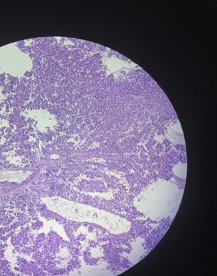
Microscopically, the tumor demonstrated:
• Pseudopapillary architecture
• Uniform polygonal cells
• Delicate fibrovascular cores
• Absence of necrosis, vascular invasion, or perineural invasion Immunohistochemistry showed:
• Nuclear β-catenin positivity
• Positive vimentin
• Positive CD10
These findings confirmed the diagnosis of SPN.
All surgical margins were negative (R0 resection). Seven regional lymph nodes were free of tumor.
The postoperative course was uneventful. The patient remains disease-free 28 months postoperatively (Figure 3).
Clinical discussion
SPN is a rare pancreatic tumor characterized by low malignant potential and excellent prognosis [3,4].
Epidemiology
A systematic review by Papavramidis and Papavramidis analyzed 718 cases and confirmed the strong female predominance and favorable survival rates [3]. A more recent systematic review by Law et al. reported increasing recognition of SPN worldwide [6].
Diagnosis
Preoperative diagnosis remains challenging due to nonspecific symptoms and normal tumor markers [8]. Imaging plays a central role, typically revealing:
• Well-circumscribed lesions
• Mixed solid and cystic components
• Peripheral calcifications
• Hemorrhagic degeneration
MRI is particularly useful in identifying hemorrhagic components and capsule integrity [9].
Definitive diagnosis relies on histopathology and immunohistochemistry. Nuclear β-catenin expression due to CTNNB1 mutation is characteristic of SPN [10].
Treatment
Complete surgical resection is the treatment of choice [3,4,11]. Even in cases with limited metastasis, aggressive surgical management is recommended [4] (Figure 2).
The recurrence rate after R0 resection ranges between 3% and 9% [3]. Five-year survival exceeds 95% [4].
Adjuvant chemotherapy and radiotherapy have limited evidence and are generally reserved for unresectable or recurrent disease [11].
Conclusion
SPN is a rare pancreatic tumor that predominantly affects adolescent and young adult females. Although classified as malignant, it demonstrates low aggressive potential. Complete surgical resection offers excellent long-term survival. Early recognition and appropriate management are essential for optimal outcomes.
References
- Frantz VK. Tumors of the pancreas. In: Atlas of tumor pathology. Washington (DC): Armed Forces Institute of Pathology. 1959: 32-33.
- Klöppel G, Solcia E, Longnecker DS, et al. Histological typing of tumours of the exocrine pancreas. WHO international histological classification of tumours. 2nd ed. Berlin: Springer. 1996.
- Papavramidis T, Papavramidis S. Solid pseudopapillary tumors of the pancreas: review of 718 patients. J Am Coll Surg. 2005; 200: 965-972.
- de Castro SM, Singhal D, Aronson DC, et al. Management of solid-pseudopapillary neoplasms of the pancreas. World J Surg. 2007; 31: 1130-1135.
- Tang LH, Aydin H, Brennan MF, Klimstra DS. Clinically aggressive solid pseudopapillary neoplasms of the pancreas. Am J Surg Pathol. 2005; 29: 512-519.
- Law JK, Ahmed A, Singh VK, et al. A systematic review of solid-pseudopapillary neoplasms: are these rare lesions? Pancreas. 2014; 43: 331-337.
- Torres OJM, Rezende MB, De Waechter FL, et al. Pancreatoduodenectomy for solid pseudopapillary neoplasm of the pancreas. Arq Bras Cir Dig. 2019; 32: e1442.
- Gurzu S, Bara T, Sincu M, et al. Solid pseudopapillary neoplasm of the pancreas: two case reports. Medicine (Baltimore). 2019; 98: e16455.
- Buetow PC, Buck JL, Pantongrag-Brown L, et al. Solid and papillary epithelial neoplasm of the pancreas: imaging features. Radiology. 1996; 199: 707-711.
- Tanaka Y, Kato K, Notohara K, et al. Frequent beta-catenin mutation and cytoplasmic/nuclear accumulation in solid-pseudopapillary neoplasm of the pancreas. Am J Pathol. 2001; 158: 1615-1622.
- Martin RC, Klimstra DS, Brennan MF, Conlon KC. Solid-pseudopapillary tumor of the pancreas: a surgical enigma? Ann Surg Oncol. 2002; 9: 35-40.